Facial Aesthetics: Concepts and Clinical Diagnosis (book)
This page contains the table of contents from the book Facial Aesthetics: Concepts and Clinical Diagnosis by Farhad B. Naini as an overview for facial aesthetics.
Part II: Clinical Diagnosis[edit | edit source]
Section I[edit | edit source]
Chapter 7: Cephalometry and Cephalometric Analysis[edit | edit source]
Natural Head Position foreword here
Hard tissue lateral cephalometric (skeletal) landmarks[edit | edit source]
Anterior facial landmarks (from superior to inferior)
- Glabella
- Nason
- Frontonasomaxillary point (abbreviated FNM or M-point)
- Orbitale
- Anterior nasal spine
- A-point
- Prosthion
- Infradentale
- B-point
- D-point
- Pogonion
- Gnathion
- Menton
Posterior facial landmarks (approximately from superior to inferior)
- Sella entrance point
- Sella
- Posterior clinoid process point
- Porion
- Condylion
- Condylar midpoint
- Articulare
- Basion
- Opisthion
- Bolton point
- Pterygomaxillare superius
- Pterygomaxillare
- Posterior nasal spine
- Gonion
- Hyoid point
Hard tissue lateral cephalometric (dental) landmarks[edit | edit source]
- Centroid
- Incision superius apicalis
- Incision superius incisalis
- Incision inferior incisalis
- Incision inferior apicalis
- Mx6.mbc
- Mn6.mbc
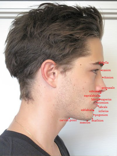
Soft tissue lateral cephalometric landmarks[edit | edit source]
- Glabella
- Nasion
- Rhinion
- Pronasale
- Subnasale
- Soft tissue A-point
- Labrale superius
- Stomion superius
- Stomion
- Stomion inferius
- Labrale inferius
- Soft tissue B-point
- Pogonion
- Gnathion
- Menton
- Cervical point
Hard tissue lateral cephalometric reference planes[edit | edit source]
Horizontal reference planes
- Sella entrance to nasion (Se-N) plane
- Delaire horizontal line (superior cranial base line)
- Sella-nasion plane
- Optic plane
- Frankfort horizontal plane
- Constructed Frankfort horizontal plane
- Maxillary plane
- Occlusal plane
- Mandibular plane
Vertical reference planes
- Nasion perpendicular
- Facial plane (skeletal)
- Delaire vertical line
- A-pog line
- Y-axis
- Facial depth line
- Posterior ramus plane
Soft tissue lateral cephalometric reference planes[edit | edit source]
- E-line
- Facial plane (soft tissue)
- H-line
- Rees aesthetic plane
- Riedel plane
- S-line
- Sn-pog' line
- Zero-degree meridian
- Z-line
Hard tissue posteroanterior cephalometric landmarks[edit | edit source]
Midline landmarks
- Crista galli
- Anterior nasal spine
- Incision superius frontale
- Incision inferius frontale
- Menton
Bilateral landmarks
- Eurion
- Latero-orbitale
- Medio-orbitale
- Zygion
- Maxillare
- Mx6.bs
- Mn6.bs
- Mastoidale
- Mental foramen
- Gonion
- Antegonion
Hard tissue posteroanterior cephalometric reference planes[edit | edit source]
Vertical reference planes
- Midsaggital plane
Horizontal reference planes
- Superior transorbital plane
- Inter latero-orbitale plane
- Inter medio-orbitale plane
- Transpetrous plane
- Interzygomatic plane
- Inferior transorbital plane
- Intermaxillare plane
- Occlusal plane
- Canine line
- Incisal line
- Intergonial (bigonial) plane
- Inter mental foramen plane
- Inferior chin plane
Linear cephalometric measurements and normative values[edit | edit source]
- Anterior facial height
- Midface height (N-ANS)
- Midface height (N perpendicular to maxillary plane)
- Lower face height (ANS-Me)
- Lower face height (Me perpendicular to maxillary plane)
- Sella-point A
- Sella-gnathion (S-Gn)
- Sella-PNS
- Posterior facial height (S-Go)
- Lower posterior facial height (Ar-Go)
Angular cephalometric measurements and normative values[edit | edit source]
- N-S-Gn
- SN to Frankfort plane
- SN to maxillary plane
- SN to functional occlusal plane
- SN to mandibular plane
- SN to posterior ramus plane
- Frankfort-mandibular plane angle
- Maxillary-mandibular plane angle
Saggital skeletal relationships[edit | edit source]
Saggital dentoalveolar relationships[edit | edit source]
Vertical skeletal relationships[edit | edit source]
Vertical dentoalveolar relationships[edit | edit source]
Inclination of the occlusal plane Anterior maxillary dental height Posterior maxillary dental height Anterior mandibular dental height Posterior mandibular dental height
Section II[edit | edit source]
Chapter 8: Facial Type[edit | edit source]
Cephalic indices[edit | edit source]
- Dolichocephalic
- Mesocephalic
- Brachycephalic
Chapter 9: Facial Proportions[edit | edit source]
Vertical facial proportions[edit | edit source]
Transverse facial proportions[edit | edit source]
Chapter 10: Facial Symmetry and Asymmetry[edit | edit source]
Classification of facial asymmetry[edit | edit source]
- Malformation
- Deformation
- Disruption
- Bilateral symmetry
- Mandibular lateral displacement
Section III[edit | edit source]
Upper face analysis[edit | edit source]
Chapter 11: The Forehead[edit | edit source]
Forehead aesthetic unit[edit | edit source]
- Superior forehead subunit
- Supraorbital ridge-lateral orbit rim subunit (inc. glabellar)
Forehead anatomy[edit | edit source]
- Squamous part of the frontal bone
- Glabellar region
- Supraorbital ridge
- Supraorbital rim
- Frontonasal suture
- Frontomaxillary suture
- Frontozygomatic suture
Frontal view
- Forehead width
Bizygomatic distance should be the widest part of the face. Bitemporal distance should be between 80-80% of bizygomatic width. Bigonial width 70-75% of bizygomatic width
- Forehead height
Profile view
- Forehead inclination
- Mildly posterior - (sloping backwards)
- Vertical
- Anterior - (opposite of men)
- Supraorbital rim projection
- Morphology of the glabellar-nasal radix region
Superior view Curvilinear relationships
Sexual dimorphism of the forehead[edit | edit source]
Supraorbital ridge is more pronounced in male's than females' Men usually have mildly posterior forehead inclination, whereas females usually have an anterior inclination Men usually have a longer forehead
Chapter 12: The Orbital Region[edit | edit source]
In Chapter 12, Dr. Farhad argues that this region, is quite possibly, the most important of all features in the face.
Orbital region subunit[edit | edit source]
- Visible eye
- Eyelids
- Superior palpebral fold
- Inferior tarsal portion
- Superior septal portion
- Superior palpebral fold
- Eyebrow
- Female eyebrow
- Male eyebrow
The orbit
- Superior orbital rim
- Supraorbital foramen
- Frontomaxillary suture
- Medial orbital rim
- Inferior orbital rim
- Infraorbital foramen
- Zygomaticomaxillary suture
- Lateral orbital rim
- Frontozygomatic suture
- Orbital part of the frontal bone
- Lacrimal bone
- Orbital part of the maxilla
- Orbital plate of ethmoid bone
- Optic canal
Eyelid anatomy
- Orbital septum
- Lacrimal gland
- Levator palpebrae superioris muscle and tendon
- Lacrimal sac
- Superior tarsal plate
- Palpebral fissure
- Inferior tarsal plate
- Medial palpebral ligament
- Lateral palpebral ligament
Surface features
- Superior orbital fold
- Upper eyelid
- Medial canthus
- Sclera with overlying conjunctiva
- Iris
- Pupil
- Lower eyelid
- Lateral canthus
- Medial limbus
- Lateral limbus
Midfacial analysis[edit | edit source]
Chapter 13: The Ears[edit | edit source]
Auricle (external ear) anatomy
- Helix
- Crus of the helix
- Auricular tubercle
- Antihelix
- Superior crus of the helix
- Inferior crus of the helix
- Triangular fossa
- Scaphoid fossa
- Concha of the auricle
- External acoustic meatus
- Tragus
- Antitragus
- Intertragic notch
- Lobule (ear lobes)
Chapter 14: The Nose[edit | edit source]
Bony skeleton of the external nose
- Nasal bone
- Frontal process of the maxilla
- Nasal process of the frontal bone
Cartilaginous skeleton of the external nose
- Septal cartilage
- Upper lateral (nasal) cartilage (paired)
- Major alar cartilage (paired)
Nasal aesthetic subunits
- Nasal radix
- Nasal dorsum
- Nasal sidewalls
- Nasal lobule
- Nasal ala
- Nasal facets
- Columella
Nasal type Hyperleptorrhine (excessively tall and narrow) Leptorrhine (tall and narrow) Mesorrhine (medium) Platyrrhine (broad and flat) Hyperleptorrhine (excessively broad and flat)
Chapter 15: The Malar Region[edit | edit source]
Anatomy of the zygomatic arch
- Frontozygomatic (FZ) suture
- Frontal process of the zygomatic bone
- Zygomatic process of the maxilla
- Zygomaticomaxillary suture
- Zygomatic arch
- Temporal process of the zygomatic bone
- Zygomatic process of the temporal bone
- Zygomatic minor
- Zygomatic minor
Chapter 16: The Maxilla and Midface[edit | edit source]
Terms used to describe position of the jaws in the saggital plane
- Prognathic
- Retrognathic
- 'Relative' prognathism
- 'Relative' retronathism
- Protrusion
- Retrusion
- Dentoalveolar protrusion
Terms of maxillary position in the vertical plane
- Vertical maxillary excess
- Vertical maxillary deficiency
Terms of jaw size
- Macrognathia
- Micrognathia
- Hypoplasia
Terms of maxillary bodily movement in the three planes of space
- Saggital plane (forward and backward)
- Vertical plane (upward and downward)
- Transverse plane (right and left)
Terms of maxillary rotation around the three axes of rotation
- Rotation around the vertical axis
- Rotation around the saggital axis
- Rotation around the transverse axis
Maxilla and midface (frontal view)
- Frontal process
- Inferior orbital margin
- Infraorbital foramen
- Zygomatic process
- Zygomaticomaxillary suture
- Alveolar process
- Canine fossa
- Canine eminence
- Incisive fossa
- Anterior nasal spine
Maxillary processes
- Zygomatic process
- Frontal process
- Palatine process
- Alveolar process
Sagittal midfacial-maxillary evaluation[edit | edit source]
Soft tissue evaluation
- Scleral exposure
(usually caused by saggital upper midfacial deficiency due to retrusion of inferior orbital rim)
- Inferior orbital rim projection
An important indicator of midfacial retrusion is reduced saggital projection of the inferior orbital rim.
- Cheek morphology and contour
- Borders of the cheek
- Superiorly: inferior orbital rim and zygomatic arch
- Inferiorly: just above the inferior border of the mandible
- Laterally: just anterior to the preauricular crease
- Medially: middle and lower third of the lateral border of the nose and nasolabial fold
- Borders of the cheek
- Midfacial curvilinear relationship
- Paranasal region
- Nasal base support
- Upper lip prominence
- Upper lip inclination
- Upper lip inclination to nasal-perpendicular
- Nasolabial angle
- Upper lip support
- True vertical plane through glabella
Dento-skeletal evaluation Transverse maxillary evaluation
When it comes to maxillary shortcomings, they can be categorised as either a:
Maxillary deficiency[edit | edit source]
Maxillary deficiency means that the maxilla is either positioned too far posteriorly or superiorly and is too narrow (which is termed specifically, maxillary hypoplasia). Deficiencies may only be the maxilla and lower midface but can involve the upper midface. If there is higher maxillary and upper midface deficiency, there may be flatness of the malar region, infraorbital rims, subpupil-mid cheek region and paranasal region.
Saggital maxillary deficiency Saggital maxillary deficiency is characterised by, but not always as
- Retrusive appearane of the upper lip
- Prognathic appearance of the maxillary due to the retrognathic nature of the maxilla
- with concomitant vertical maxillary deficiency (hereon, VMD) leading upward and forward mandibular rotation and increased chin prominence insomuch as to cause over-closure of the lips which causes them to curl together and outwardly.
- Reduced or negative facial contour angle (concave midfacial appearance)
- Increased nasolabial angle (lower component) due to the posterior inclination of the upper lip, though the upper component may be negative, as the nasal tip is often hanging
- Nasal prominence due to retrusive upper lip and lower midface and paranasal hollowing
- Maxillary incisor exposure reduced or non-existent (even when smiling in severe cases) with concomitant VMD the maxillary incisor tips tend to be at a vertical level superior to the upper lip
- Retrusion of the anterior wall of the maxillary sinus resulting from retroposition or hypoplasia of the maxilla.
- Dental occlusion
- Reverse incisor overjet
- Tendency to posterior crossbite
- Dentoalveolar compensation due to skeletal pattern (proclination of maxillary incisors, with or without retroclination of the mandibular incisors)
- Chin-neck (submental) length is normal
Vertical maxillary deficiency
- Reduced total anterior facial height, square face in frontal view
- Lack of maxillary skeletal development, no bone between the root of the teeth and the nasal floor, sometimes the roots can extent to the maxillary sinuses
- Upper lip incisor relationship
- Reduced exposure of the maxillary incisors in relation to the upper lip in repose
- Reduced or no teeth showing upon a smile
- Anterior mandibular rotation (having an affect on saggital prominence of chin, deep mentolabial fold, reduced mentolabial angle)
- Reduced mandibular plane angle
- Bigonial width is increased, the gonial angles are reduced
- Lips are squared together.
- Mouth width is increased; oral commisures turn downwardly
- Nose tends to have an increased alar base width
- Palatal vault tends to be flat and broad
- Dental occlusion
- Incisor overbite
- Free way space increased to 10mm
Transverse maxillary deficiency
- Relative transverse maxillary deficiency
- Absolute transverse maxillary deficiency
(usually a result of true skeletal narrowing of the maxilla)
Maxillary excess[edit | edit source]
Maxillary excess is overdevelopment on the maxilla, which can be present in the saggital, vertical and transverse planes.
Saggital maxillary exesss
Vertical maxillary excess
- Increased total anterior facial height
- Excessive inferior maxillary skeletal development
- Total VME
- Posterior VME
- Anterior VME
- Upper lip-incisor relationship
- Increased exposure of the maxilary incisors
- Increased dentogingival exposure upon smiling
- Posterior (downward and backward) mandibular rotation
- Increased mandibular plane angle
- Lip incompetence
- Nose
- Palatal vault
- Incisor overbite
Transverse maxillary excess
Maxillary asymmetry[edit | edit source]
Lower facial analysis[edit | edit source]
Chapter 17: The Lips[edit | edit source]
- Philtrum
- Philtrum ridges/columns
- Cupid's bow
- High points of the vermillion
- White roll
- Upper lip vermillion
- Upper lip tubercle
- Vermillion border
- Lower lip vermillion
- Oral commissures
- Nasolabial groove
- Mentolabial groove
Lip morphology[edit | edit source]
Lip height Lip thickness Lip contour Lip curvature Lip curl Lip inclination
Lip posture Lip prominence
Chapter 18: Mentolabial (Labiomental) Fold[edit | edit source]
Mentolabial fold depth
Mentolabial angle
- Upper component
- Lower component
- Soft tissue factors
- Skeletal factors
- Dentoalveolar factors
Advantages of mandibular advancement over genioplasty
- Lower lip position
- Mentolabial angle
- Lower anterior facial height
- Dental occlusion
Chapter 19: The Mandible[edit | edit source]
- Condylar process
- Condylar head
- Condylar neck
- Pterygold fovea
- Coronoid process
- Ramus
- Oblique line
- Gonial angle
- Alveolar process
- Body (mandibular corpus)
- Mental foramen
- Inferior border
- Mental protuberance
- Mandibular foramen
- Superior and inferior mental spines
- Sigmoid notch
Mandibular subunits
- Condylar subunit
- Coronoid subunit
- Ramus
- Gonial angle
- Body
- Dentoalveolar subunit
- Inferior border
- Mandibular symphysis
Mandibular growth rotations
- Anterior rotation
- Posterior rotation
Bjork's seven structural signs of mandibular growth
- Inclination of the condylar head
- Curvature of the mandibular canal
- Shape of the lower border of the mandible
- Inclination of the mandibular symphysis
- Interincisal angle
- Intermolar and interpremolar angle
- Lower anterior facial height
Chapter 20: The Chin[edit | edit source]
Chin excess and deficiency[edit | edit source]
- Primary progenia (primary sagittal chin excess):
- Horizontal osseous macrogenia
- Increased thickness of the chin pad
- Combination of above
- Retrogenia
- Primary retrogenia
- Secondary retrogenia
Chapter 21: Submental-Cervical Region[edit | edit source]
Submental anatomy in relation to the mandible
- Mylohyoid muscle
- Submandibular salivary gland
- Genloglossus muscle
- Genlohyoid muscle
- Anterior belly of digastric muscle
- Medial pterygoid muscle
Section IV[edit | edit source]
- Teeth and the alveolar bone make up the dentoalveolar process. Loss of teeth will case absorption of the alveolar process and thus cause a reduction in lower anterior facial height.
Chapter 22: Dental-Occlusal Relationships: Terminology, Description and Classification.[edit | edit source]
Chapter 22 covers the importance of the relationships between the dental-occlusal relationships together with the relationship of the dento-alveolar process and the craniofacial complex. The chapter begins by refreshing your knowledge on dental anatomy and terminology, discusses different kinds of malocclusion and deformities and ideal ratios.
Chapter 23: Smile Aesthetics[edit | edit source]
There are two types of smiles
- Posed smile
A fake smile.
- Spontaneous smile
Regarded as involuntary, linked to the emotions, considered a 'genuine' smile. Also called a Duchenne smile.
- Stages of smile formation
- Stage I:
- Stage II:
Chapter 24: Dentogingival Aesthetics[edit | edit source]
Dentogingival complex is divided into two subunits which form one aesthetic unit of the face.
- Gingival subunit
- Dental subunit
Dental aesthetics[edit | edit source]
Tooth shape is genetically determined and subject to individual variability, each individual will bit into one of three spectra.
- Ovoid
- Rectangular
- Triangular
Many theories on ideal tooth-shape have been discussed.
- Tooth shape-face shape correlation
- Tooth width-face width correlation
- SAP theory
- Sex
- Age
- Personality
Dental arch form
- Zone of soft tissue equilibrium: the position of stability of tongue, lips and cheek musculature.
Orthodontist Allan G Brodie described the relationship between orofacial musculature and tooth position, it was vertified by Weinstein et al., who concluded that "teeth are maintained in a state of equilibrium between the perioral soft tissue forces"
- Ovoid arch form
- Square arch form
- Tapered arch form
